Elderly mid 80s pt with weeks of worsening SOB and SOBOE. Now SOB on walking to bathroom. 6 months ago doing own shopping. Orthopnoea + PND. HR 30-50 AF with slow ventricular response. On Metoprolol 12.5mg BD and Apixaban. Awaiting public cardiology review.
TTE ?Valvular Heart disease / Interstitial Syndrome.
MV look a bit thickened and stiff. AV doesnt look too bad. Aortic Root appears mildly dilated.
That Aortic valve really doesnt look too bad. Trileaflet, coming together nicely.
Was attempting to align the image for Aortic PWD Trace to calculate CO. Marked turbulence seen. Further look on PSAX showed this:
You could see the turbulent flow originating from the edges of the posterior leaflet.
There was significant valvular incompetance noted in the other valves as well:
MR on PLAX. Does not look so bad does it?
You could see where the MR is originating on PSAX:
Corroborating with SC and A4C (ignore the ‘tricuspid’ label please)
MR is really moderate to severe, as it touches the top of the atrium.
Next, looking at the tricuspid:
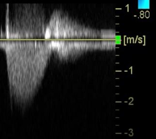
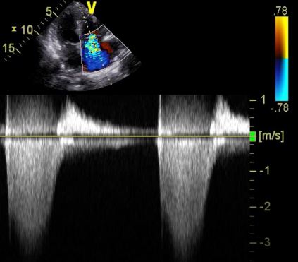
TRVMax on this view was 2. That’s not too bad. Or is it?

Below is a flipped view of the heart (TV on the right of screen). The alignment of the probe is more inline with the TR Jet.

Here we appreciate the importance of the doppler plane being inline with the flow of blood.
The TR Jet was so strong blood was flowing up the hepatic veins. Also note the veins are more prominent than usual.
Finally, the valve less commonly seen on TTE: The Pulmonary Valve. This is a zoomed in view of the top right part of the PSAX window. The elusive PV slips into view briefly as the patient breathes.
Trivial PR noted (red bits).
This patient had no interstitial syndrome (B lines / effusions) on her lung windows. This suggests the primary pathology is related to valvular heart disease and the structural heart abnormalities that result from it. It could also be iatrogenic (pt’s on metoprolol) given the history.
Lesson: Multiple views from at least two planes are needed to make a clear diagnosis of pathology given the fact that each US window is really a 2 dimensional plane that may not accurately reflect reality.
cover image from pexels.com (creative commons)

